HFO detection in SEEG: Recent insights from the research community
High-frequency oscillations (HFOs) have been increasingly investigated for their role in seizure onset localization and clinical outcome prediction. Below, we shed light on recent research studies that provide major insights on automated HFO detection.
Combining interictal biomarker features to predict the epileptogenic zone
Comment on Partamian H. et al., Predicting surgical outcome in drug-resistant epilepsy by combining interictal biomarkers within a machine learning framework.(2026) Sci. Reports.
In this study, the authors explore the potential of interictal biomarkers, and more specifically spikes and ripples, as markers of the epileptogenic zone. A major strength of the work lies in the characterization of these biomarkers through a broad set of temporal, spectral, and spatial features, and the subsequent comparison of the predictive power of individual vs multi-feature combination models.
Interictal iEEG recordings from 62 patients with drug-resistant epilepsy were analysed, and spike and ripple features (rate, power, propagation indices, distances, and spike–ripple co-occurrence) were combined within a Random Forest machine learning framework.
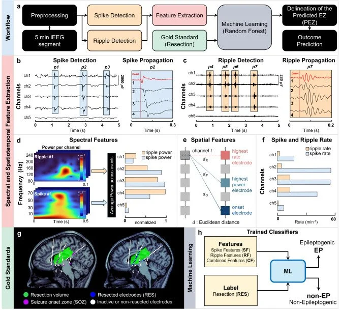
Following the comparison of several classifiers, results showed that the model including a combination of spike and ripple features achieved the best performance for predicting epileptogenic contacts, reaching an AUC-ROC of 0.89 and a 74% overlap with the resected tissue. Interestingly, while combining several features offered a clear advantage over individual feature predictions, the contribution of ripple features remained modest.
These findings support the idea that no single biomarker feature fully captures the complexity of the epileptogenic network and that integrating information from multiple interictal biomarkers can provide a more robust estimate of epileptogenic tissue. Future work should consider extending such a multimodal framework to include fast ripples, which are often considered more specific markers of epileptogenic tissue than ripples.
Illustration taken from the publication Partamian H., et al. (2026), Sci Reports.
Full article: Partamian H., et al. (2026). Sci Reports;16:15166.
Other interesting recent articles on HFOs detection
Fast ripples: a clinical validation study.
Comment on Nevalainen, P. et al., Fast Ripples Measured From Overnight SEEG Recordings as Markers of the Epileptogenic Zone: A Multicenter Validation Study (2026). Neurology.
In their study, Nevalainen and colleagues assessed the impact of removing fast ripples, spikes, or fast ripples co-occurring with spikes, on the surgery outcome, in a retrospective dataset of 73 patients from 4 epileptic centers. They calculated the resection ratios of the detected events by determining resected SEEG contacts.
They found that a high resection ratio of fast ripples associates with good post-surgical outcome in a greater proportion when the reseaction ratio is greater than 60%. This makes fast-ripples a good predictor of post-surgical outcome, similarly when they occur alone or when they co-occur with spikes. A high resection ratio of spikes alone was, however, not a good predictor of post-surgical outcome.
Full article: Nevalainen, P., et al. (2026). Neurology; 106(2), e214511.
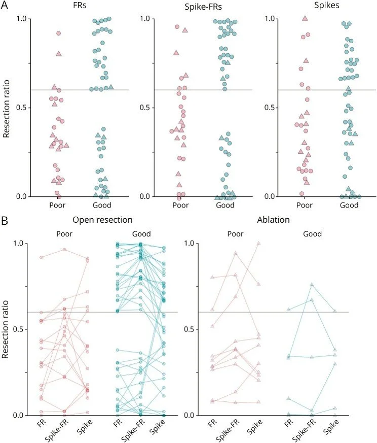
Illustration taken from the publication Nevalainen, P., et al (2026), Neurology.
Automatic detection of artefactual false positive HFOs
Comment on Tan S.B. et al., A comprehensive, physician-trained algorithm to remove artifactual false positive high frequency oscillations in long-term intracranial EEG (2026). Journal of Neural Engineering.
Dr Stacey’s team at University of Michigan developed a detector for artefactual HFOs, called the “Michigan Intracranial Artifact Filter (MIAF)”. They trained it and tested it on iEEG signal dataset where HFOs were detected both by their automatic HFO detector as well as by expert neurologists. HFOs classified as non-artefacts alone correlated better with the seizure onset zone and resected volume than HFOs before artifact removal, demonstrating that the artefacts were simply adding noise to the signal.
The use of the MIAF tool on iEEG signal improves clinical utility of HFOs, increasing their predictive value. Interestingly, these results may apply to other detections such as spike and seizure biomarkers.
Full article: Tan S.B., et al. (2026). J. Neural Eng; 23 026019.
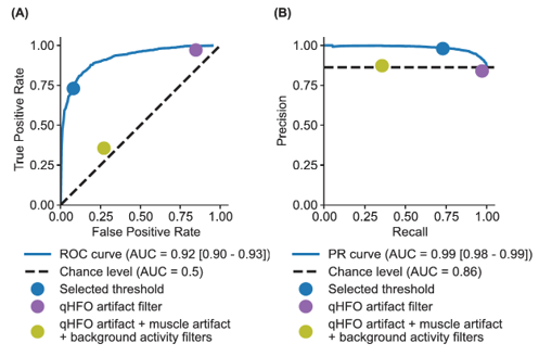
Illustration taken from the publication Tan, S.B., et al., J. Neural Eng, 2026.